Posts

Running Injuries

Image
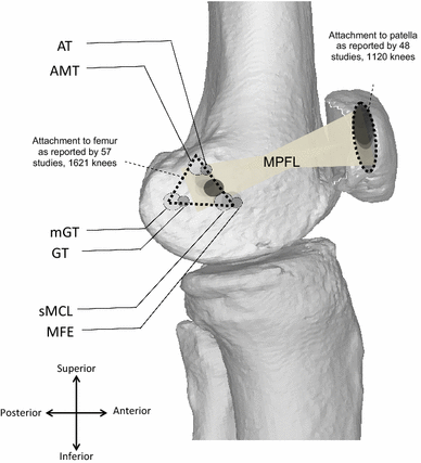
Running Injuries Running is one of the most common forms of exercise. It is a convenient and efficient workout that can be an escape from the noise and stress of the outside world; it can be a great way to get your day started off on a good foot, or to decrease stress after a long day of work.  Many runners use running as their primary, and sometimes only source of physical activity.  Running places a lot of stress on our body and just like with any sport there are inherent injury risks.  When compared to other types of aerobic exercise such as biking, swimming, or walking, running has a higher risk of injury.  This is tied to the amount of impact that our body undergoes during running and the need to dissipate those forces between muscles and joints.  In this blog we will discuss some common running injuries, some potential causes, and some potential management strategies. Common running injuries According to Francis et al.  the knee is the most c...

Article Summary: Effectiveness of Active Rehabilitation Program on Sports Hernia: Randomized Control Trial

Image
Effectiveness of Active Rehabilitation Program on Sports Hernia: Randomized Control Trial Objective:  "To determine whether an active rehabilitation program that involves repetitive effortful muscle contractions, including core stability, balancing exercises, progressive resistance exercises, and running activities, after a sports hernia, is effective." Population:  40 male soccer players age 18-25 with groin pain with sport for at least 2 months. Methods:  Study Design: Single Blind RCT Group A: Active Rehabilitation Program (also received Group B intervention) Group B: Conventional Treatment Program Randomization via envelope method.  Staff therapist drew an envelope and handed to treating therapist. Both groups instructed to perform stretching exercises for adductor, hamstring, and hip-flexor musculature on days between treatment days. Did not specify if there were different treating therapists. Did not specify any training to provide c...

Open Kinetic Chain Knee Extension After Articular Cartilage Repair in the Knee: Is it Safe?

Image
Open Kinetic Chain Exercise After Articular Cartilage Repair in the Knee: Is it Safe? After articular cartilage repair in the knee, specifically with the tibiofemoral joint, there is a period of protected weight bearing.  Early post-operatively we know that regaining range of motion, regaining quadriceps activation, and decreasing excess inflammation and swelling is important.  Commonly weight bearing is progressed to full by about 8 weeks, sometimes a little longer or shorter depending on the surgeon, the surgery, and lesion factors.   Deficits in quadriceps strength are seen far beyond discharge from rehabilitation and have been seen up to 7 years post op.  Knowing this, it is easy to want to get after quadriceps strengthening early.  There has been a lot of talk in the physical therapy community about "underdosing" our patients when we claim that we are "strengthening" them.  We are aware that we should be cautious with weight bearing exerc...

Article Summary: Post-Operative Sport Participation and Satisfaction with Return to Activity After Matrix-Induced Autologous Chondrocyte Implantation in the Knee

Post-Operative Sport Participation and Satisfaction with Return to Activity After Matrix-Induced Autologous Chondrocyte Implantation in the Knee Purpose:  To investigate:  The level and improvement in activity in patients at two years after matrix-induced autologous chondrocyte implantation (MACI),  What factors are associated with post-operative (and improvement in) activity level, and  Whether patients are satisfied with their ability to participate in recreational and/or sporting activities." Methods: Study Design:  Prospective Cohort (level of evidence: 3) Population:  150 patients that underwent MACI (Started with 160 but 10 were unable to be found for 2 year follow up so were excluded) 83 tibiofemoral lesions 63 medial femoral condyle 20 lateral femoral condyle 67 patellofemoral lesions 35 patella 32 trochlea 92 patients had undergone prior surgery, most commonly arthroscopy and meniscectomy (88 arthroscopy,...

Runner's Knee: Patellofemoral Pain - Causes and Treatment Strategies.

Anterior Knee Pain in the Active Individual: Patellofemoral Pain Anterior knee pain is a common complaint among that active population.  It can have many causes and can be from a few different causes.  The two most common in young, active individuals are patellofemoral pain, and patellar tendinopathy.  In this post, we will be talking about patellofemoral pain. Patellofemoral pain: What is patellofemoral pain? Also referred to as "Runner's Knee" Your knee cap (patella) rests within a groove at the bottom of your femur.  This groove is referred to as your trochlea.  As our knee bends and straightens, our patella glides along this trochlea.  In our body, our quadriceps muscle becomes our quadriceps tendon who's fibers house the patella and continue below to form our patellar tendon which attaches onto our tibia (shin bone).  The patella serves to increase the mechanical advantage of our quadriceps allowing it to produce much more force than...

The Basics of Concussion Rehab

Image
What is Concussion Rehabilitation? What is a concussion? To answer that question I think it is important to first understand what a concussion is not. A concussion is not a structural injury, meaning that it will not show up using any of our traditional imaging techniques (MRI and CT scan). If you or someone you know initially went to the ER after suffering a head injury and imaging was ordered, it was not to “rule in” a concussion, it was to “rule out” other, more serious pathology such as a brain bleed. So if it is not a structural injury what is it? Rather than impact causing the injury, rapid movement causes shearing forces that cause damage to parts of our neural cells called “axons.” This disruption causes a change in ion concentration inside and outside the cell as well as a spike in glutamate, our body’s main excitatory neurotransmitter. Initially, this change causes many cells to fire in what is called “spreading depolarization” and a lot of energy is releas...

Evaluation and Treatment of the Medial Patellofemoral Ligament (MPFL)

Image
Evaluation and Treatment of the Medial Patellofemoral Ligament (MPFL) Basic Facts: 2-3% of all knee injuries 15-44% recurrence; 49% in those that have previously dislocated twice. ~ 16% re-dislocation in those treated non-op; ~ 33% have residual Sx; remaining 50% will be asymptomatic. Anatomy: Origin: superior medial aspect of the patella Insertion: space b/n adductor tubercle and medial epicondyle Some fibers blend with VMO Function: As knee flexes, VMO and MPFL pull patella medially to help it remain in trochlea Provides stability against lateral translation Biomechanics: Provides 50-60% restraint to lateral patellar translation at 0-30 degrees flexion If sectioned, patella displaces laterally even w/ all other medial stabilizers intact Loses tension as the knee is flexed, tightest b/n 25-30 degrees of knee flexion and creates a medial shift; this shift is not appreciated in knees w/ resected MPFL. Patella enters the trochlea around 15-20 degrees of knee...